PARASITES IN THE EYE: THE PARASITIC INFECTIONS THAT CAN THREATEN VISION
The eye is a highly specialized organ with delicate tissues that can be damaged by inflammation, infection and direct invasion. Although relatively uncommon, several parasites can involve the eye, affecting the cornea, retina, choroid, optic nerve, eyelids or surrounding tissues. Some infections can cause permanent visual impairment when inflammation or structural damage involves critical areas of the visual system.
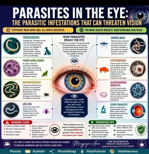
HOW CAN PARASITES REACH THE EYE? Parasites can reach ocular tissues in several ways. Some larvae migrate through the bloodstream after entering the body, while others are transmitted by insects or contaminate food, water, soil or surfaces. In certain infections, the organism directly enters or colonizes ocular tissues. The route of infection often determines which part of the eye is affected.
TOXOCARIASIS: WHEN ROUNDWORM LARVAE REACH THE EYE One of the best-known parasitic eye infections is ocular toxocariasis, caused by the larvae of Toxocara canis or Toxocara cati, roundworms associated primarily with dogs and cats. Humans can accidentally ingest microscopic eggs from contaminated soil, hands or other environments. After hatching, larvae can migrate through tissues and occasionally reach the eye.
OCULAR LARVA MIGRANS The eye can become a destination for a migrating Toxocara larva. The resulting inflammatory response may produce a granuloma involving the retina or surrounding tissues. Ocular toxocariasis commonly affects one eye and can cause reduced vision, floaters, light sensitivity, eye inflammation and, in severe cases, substantial loss of vision.
TOXOPLASMA GONDII: A MAJOR CAUSE OF INFECTIOUS RETINAL INFLAMMATION Toxoplasma gondii is a microscopic protozoan parasite capable of producing ocular disease. Ocular toxoplasmosis can cause necrotizing retinitis, inflammation and damage to retinal tissue. A characteristic active lesion may appear as a focal area of white retinal inflammation surrounded by vitreous inflammation.
WHY TOXOPLASMOSIS CAN BE SERIOUS The retina contains highly specialized cells responsible for converting light into neural signals. Damage to the retina can therefore have lasting consequences, particularly when an active lesion involves the macula, optic nerve or other visually important structures. Scarring may remain after an episode has healed, and recurrent disease can occur.
CONGENITAL TOXOPLASMOSIS AND THE EYE Toxoplasma infection acquired during pregnancy can affect the developing fetus. Ocular disease may become apparent at birth or much later in life, meaning congenital infection can have long-term ophthalmological consequences. Reactivation of retinal lesions is also possible.
ONCHOCERCIASIS: THE PARASITE BEHIND RIVER BLINDNESS Onchocerca volvulus is a filarial nematode transmitted by infected blackflies. Repeated exposure to infected bites can result in the development of adult worms and microfilariae within human tissues. When microfilariae and the resulting inflammatory response involve ocular structures, progressive visual damage can occur. This is why onchocerciasis is commonly known as river blindness.
LOA LOA: THE AFRICAN EYE WORM Loa loa is another filarial parasite capable of entering ocular tissues. It is transmitted by the bite of infected Chrysops flies in parts of Central and West Africa. Adult worms can migrate through subcutaneous tissues and occasionally become visible moving across the surface of the eye.
DIROFILARIA: A WORM THAT CAN FORM OCULAR NODULES Dirofilaria species are filarial nematodes normally associated with animals. Humans can occasionally become accidental hosts following mosquito transmission. Ocular or periocular infection can present as a small nodule in the eyelid or surrounding tissue and may resemble a benign cyst or other lesion. A 2026 CDC report documented Dirofilaria repens infection presenting as an eyelid nodule in the United States.
CYSTICERCOSIS: PARASITIC CYSTS AROUND THE EYE Taenia solium, the pork tapeworm, can cause cysticercosis when larval cysts develop in human tissues. Cysts can occasionally involve the eye or tissues surrounding it. Depending on their location, ocular cysticercosis can cause inflammation, visual disturbance and mechanical complications.
ACANTHAMOEBA: A MICROSCOPIC ORGANISM THAT CAN DESTROY THE CORNEA Acanthamoeba is a free-living microscopic amoeba rather than a conventional intestinal parasite. It can cause Acanthamoeba keratitis, a serious infection of the cornea. Symptoms can include severe eye pain, redness, tearing, sensitivity to light, foreign-body sensation and reduced vision.
WHY ACANTHAMOEBA KERATITIS CAN BE DIFFICULT TO RECOGNISE Acanthamoeba keratitis can initially resemble other forms of corneal infection. Delayed recognition may allow progressive corneal damage and scarring. Contact lens use, particularly when lenses or storage systems are exposed to contaminated water, is an important recognised risk factor.
THE IMMUNE RESPONSE CAN CAUSE DAMAGE Parasites do not necessarily have to physically destroy large amounts of tissue to threaten vision. The immune response directed against them can produce substantial inflammation. Within the confined structures of the eye, inflammation, swelling and tissue destruction can interfere with retinal, corneal or optic nerve function.
WARNING SIGNS THAT SHOULD NOT BE IGNORED Sudden or progressive changes in vision require prompt ophthalmological assessment. Important warning signs include:
- New or increasing floaters
- Flashes of light
- Blurred or reduced vision
- Eye pain
- Significant redness
- Marked sensitivity to light
- A new dark or missing area in the visual field
- Swelling or a persistent lump around the eye
- A visible moving organism on or around the eye
These symptoms are not specific to parasitic disease and can occur with many other eye conditions. Some causes of sudden visual loss are emergencies.
HOW OCULAR PARASITIC INFECTIONS ARE INVESTIGATED Diagnosis depends on the suspected organism and the part of the eye involved. Ophthalmologists may use slit-lamp examination, dilated retinal examination, retinal imaging, ultrasound, blood tests, antibody testing, molecular testing or examination of ocular fluid or tissue in selected cases. A negative blood test does not necessarily exclude a localized ocular infection, particularly when the organism is confined largely to the eye.
TREATMENT DEPENDS ON THE PARASITE There is no single treatment for "eye parasites." Management must be directed at the specific organism and its location. Depending on the infection, treatment may involve antiparasitic or antimicrobial medication, anti-inflammatory therapy, procedures or surgery. In ocular toxoplasmosis, for example, treatment decisions depend heavily on lesion location, severity and the risk to vision.
THE EYE IS NOT AN ISOLATED SYSTEM The presence of a parasite in the eye can reflect a wider infection elsewhere in the body. Conversely, ocular involvement may be the most obvious manifestation of an otherwise subtle infection. Understanding the parasite's complete life cycle, route of transmission and tissue migration is therefore important when investigating suspected infection.
PREVENTION STARTS WITH INTERRUPTING TRANSMISSION Basic preventive measures can reduce exposure to several parasites associated with ocular disease. These include careful hand hygiene after contact with soil or animal faeces, appropriate pet parasite control, thoroughly cooking meat, safe food and water practices, protecting against insect bites in endemic regions and following safe contact-lens hygiene. Prevention is particularly important for infections capable of causing permanent tissue damage.
THE BIGGER PICTURE Parasites affecting the eye demonstrate how complex host-parasite interactions can become. A microscopic protozoan, migrating larva, filarial worm or free-living amoeba can interact with completely different tissues and trigger very different forms of disease. Some infections remain localized, while others are manifestations of organisms migrating through the body.
The important lesson is that unusual visual symptoms should never automatically be attributed to a parasite, but neither should rare infectious causes be overlooked when exposure history, travel history, clinical findings and ocular examination point in that direction. Early identification of a sight-threatening infection can make an important difference to the outcome.
Feel free to share this article without making any changes to increase awareness and help others learn more about parasite biology.
Copyright Maryjayne Aria . All rights reserved.